Human Eye and the Sense of Sight

Sight is probably the most developed sense in humans, followed closely by hearing. The human eye is the organ of vision. It has a complex structure consisting of a transparent lens that focuses light on the retina. The retina is covered with two basic types of light-sensitive cells-rods and cones. The cone cells are sensitive to colour and are located in the part of the retina called the fovea, where the light is focused by the lens.

The rod cells are not sensitive to colour, but have greater sensitivity to light than the cone cells. These cells are located around the fovea and are responsible for peripheral vision and night vision. The eye is connected to the brain through the optic nerve. The point of this connection is called the “blind spot” because it is insensitive to light. Experiments have shown that the back of the brain maps the visual input from the eyes.

The brain combines the input of our two eyes into a single three-dimensional image. In addition, even though the image on the retina is upside-down because of the focusing action of the lens, the brain compensates and provides the right-side-up perception. Experiments have been done with subjects fitted with prisms that invert the images. The subjects go through an initial period of great confusion, but subsequently they perceive the images as right side up.

Eye structure

In a number of ways, the human eye works much like a digital camera:

  1. Light is focused primarily by the cornea, i.e. the clear front surface of the eye, which acts like a camera lens.
  2. The iris of the eye functions like the diaphragm of a camera, controlling the amount of light reaching the back of the eye by automatically adjusting the size of the pupil (aperture).
  3. The eye’s crystalline lens is located directly behind the pupil and further focuses light. Through a process calledaccommodation, this lens helps the eye automatically focus on near and approaching objects, like an autofocus cameralens.
  4. Light focused by the cornea and crystalline lens (and limited by the iris and pupil) then reaches the retina i.e. the light-sensitive inner lining of the back of the eye. The retina acts like an electronic image sensor of adigital camera, converting optical images into electronic signals. The optic nerve then transmits these signals to the visual cortex, i.e. the part of thebrainthat controls our sense of sight.
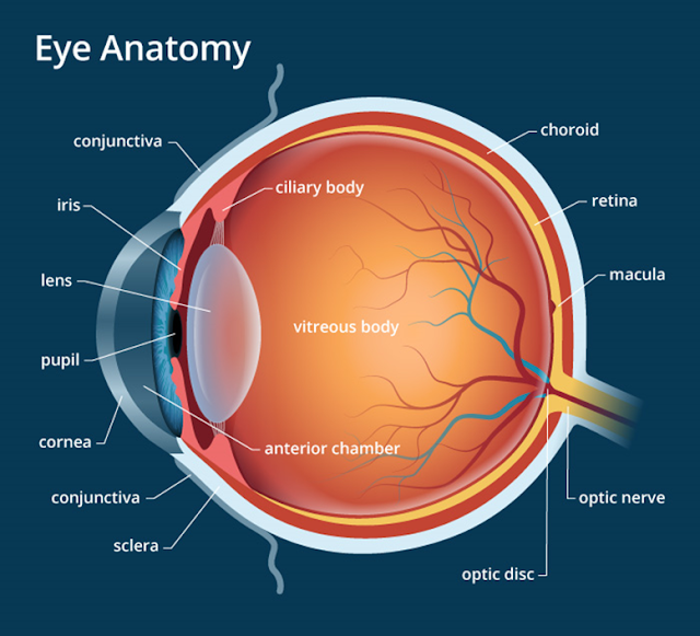
Human eye structure, organ of sight
  • Sclera:The knowledge of eye parts and functions is both useful and interesting. The white part of our eyes is known as sclera. It consists of fibrous tissues. The job of this part it to protect the internal parts of the eye.
  • Cornea:The transparent tissue through which light enters the eyes is called cornea. It exists in front of the eye. That is why, it looks prominent among different parts of the human eye.
  • Iris:Iris is made up of muscles which contract or relax in order to adjust the amount of light which enters the eye.
  • Pupil:There are many interesting facts about the pupil. It is, actually, an aperture controlled by the iris muscles. For instance, when it is dark, the iris muscles relax, causing the pupil to open up wider. Therefore, more light enters the eyes to enable us to see better. On the other hand, the pupil becomes narrow due to the contraction of the iris muscles. It occurs in the presence of excessive light in order to protect the cells of the eyes.
  • Lens:The lens is present behind the pupil. Light enters the pupil, passes through the lens and is focused on the retina of the eye. The lens is capable of changing its shape in order to help us see near or far off objects. This is about how the human eye works.
  • Retina:Digging further deeper into the human eye anatomy, you will come across the retina. The retina converts light into electrical signals which are transferred to the brain for processing. The retina contains two types of cells: rods and cones. These cells are sensitive to light. Rods are important for night-time vision when there is little light. On the other hand, cones play an important role in helping us see colours.
  • Optic Nerve:The optic nerve takes electrical signals from the retina of the eye to the brain.

Eye Function

Eye function renders you the sense of sight. They convert light into electrical signals. Afterwards, the brain deciphers these electrical signals into images. Properly working healthy eyes are able to perform the following functions.

  • It is one of the important human eye facts that they help us view our surroundings.
  • Our eyes enable us to see colours. Certain organisms, such as dogs, cannot see colours and, so, their world is black and white.
  • Eyes help us see near as well as distant objects.

Eye Problems

Colour blindness or “Daltonism” is a common abnormality in human vision that makes it impossible to differentiate colours accurately. One type of colour blindness results in the inability to distinguish red from green. This can be a real handicap for certain types of occupations. To a colour-blind person, a person with normal colour vision would appear to have extrasensory perception. However, we want to reserve the term “extrasensory perception” for perception that is beyond the range of the normal.

Conjunctivitis in the Human Eye

These extremely delicate organs can fall prey to different eye problems due to allergic factors, unhygienic conditions and the old age. Below are some of the most common diseases of eyes.

Conjunctivitis

Conjunctivitis has got a common name – the pink eye disease. It is an inflammation of conjunctiva, the transparent membrane which covers the sclera. Inflammation of tiny blood vessels found in conjunctiva makes our eyes appear red.

Cataracts

Cataracts in Human eye
Cataracts in Human eyes

Cataracts affect the eye lenses. They may occur due to old age or exist as a defect by birth. Parts of lenses become opaque, blocking the passage of light to the retina, thereby obstructing our vision.

Glaucoma

Glaucoma is a disease which leads to blindness due to a damage to the optic nerve. When the optic nerve fails to transmit electric signals to the brain, an individual is unable to see at all.

Human Heart Structure and Functions

The Human heartis a hollow, muscular organ that pumps blood through the blood vessels by repeated, rhythmic contractions.

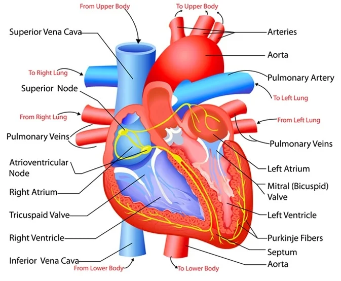
Human Heart parts
Human Heart parts

The human heart is not just the physical organ, according to the human understanding. It also holds a special place in our collective psyche as it is equated with love. It has many other associations, but here we shall just outline a few examples:

  • · have a heart – be merciful
  • · change of heart – change your mind
  • · to know something by heart – memorise something
  • · broken heart – to lose love
  • · heartfelt – deeply felt
  • · have your heart in the right place – to be kind
  • · cry your heart out – to grieve
  • · heavy heart – sadness
  • · have your heart set on – to want something badly.

Heart structure

A heart is the main organ of our body’s circulatory system, pumping blood throughout the body. it is a muscular pump which contracts at regular intervals in order to squeeze the blood through it into the blood vessels.

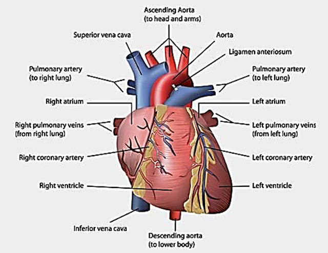
Human Heart structure

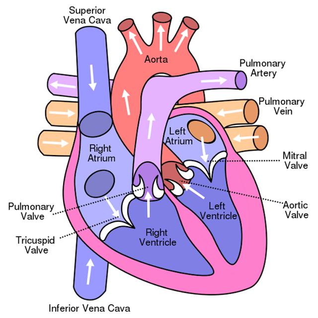
The human heart has got four chambers; two upper chambers (the atria) and two lower ones (the ventricles), which work in coordination for the successful execution of different heart functions. The right atrium and right ventricle together make up the “right heart,” and the left atrium and left ventricle make up the“left heart.” A wall of muscle calledthe septumseparates the two sides of the heart. The right chambers contain unclean or the deoxygenated blood. On the other hand, the left chambers contain clean or the oxygenated blood. Talking of the heart structure, it is made up of connective tissue and cardiac muscles. The latter is a type of involuntary muscle, that is, it contracts and relaxes on its own without requiring you to deliberately apply force. The heart is a delicate organ. Any damage to it will cause disturbance in the heart functions. So, it gets protection from the rib cage.

A double-walled sac called thepericardiumencases the heart, which serves to protect the heart and anchor it inside the chest. Between the outer layer, the parietal pericardium, and the inner layer, the serous pericardium, runs pericardial fluid, which lubricates the heart during contractions and movements of thelungsand diaphragm.

The human heart’s outer wall consists of three layers. The outermost wall layer, or epicardium, is the inner wall of thepericardium. The middle layer, ormyocardium, contains the muscle that contracts. The inner layer, orendocardium, is the lining that contacts the blood.

The tricuspid valve and the mitral valve make up the atrioventricular (AV) valves, which connect the atria and the ventricles. The pulmonary semi-lunar valve separates the right ventricle from thepulmonary artery, and the aortic valve separates the left ventricle from the aorta. The heartstrings, or chordae tendinae, anchor the valves to heart muscles. The sinoatrial node produces the electrical pulses that drive heart contractions.

Concerning shape, it looks like a cone. The base of this cone is positioned upward which gradually tapers down to the apex. The pumping organ is roughly the size of a wrist, while measuring 12 cm, 8 cm and 6 cm along length, width and thickness, respectively.

The effect of exercise on the muscles of the heart organ is the same as shown by the skeletal muscles. That is, as an outcome of exercise, they grow both in size and strength. That is why, the well-trained athletes usually have much larger hearts. Such a thing also makes the heart functions better.

The Functions of the Human Heart

The heart circulates blood through two pathways: the pulmonary circuit and the systemic circuit. The heart functions involve the supply of oxygenated blood to all parts of the body. The circulatory system consists of three main types of blood vessels: arteries, veins and capillaries.

Arteries carry oxygenated blood and distribute it to different parts of the body. On the other hand, the veins receive unclean blood, containing carbon dioxide and waste products, from the body parts and take it back to the heart.

The human heart contains both the oxygenated and deoxygenated blood. It is its job to keep both of them separate in order to avoid contamination of the pure blood. The right chambers of the heart receive blood, devoid of oxygen, from the veins. This blood then goes to thelungsto receive oxygen and get rid ofcarbon dioxide.

Afterwards, this oxygen-rich blood goes back to the heart from where it reaches all the body parts through the arteries. In this way, every individual cell is able to get oxygen. This oxygen will assist in the process of extracting energy from food.

A healthy human heart contraction happens in five stages.

  1. In the first stage (early diastole), the heart is relaxed.
  2. Then the atrium contracts (atrial systole) to push blood into the ventricle.
  3. Next, the ventricles start contracting without changing volume.
  4. Then the ventricles continue contracting while empty.
  5. Finally, the ventricles stop contracting and relax. Then the cycle repeats.

Valves prevent backflow, keeping the blood flowing in one direction through the heart.

Function of the Blood and Blood Vessels:

The function of the blood is to serve as a circulatory fluid. The blood carries the respiratory gases and nutrients to every individual cell in the body. On its return, it brings carbon dioxide, a waste respiratory gas, for its discharge out of the body.

Diseases of the human heart

Coronary Artery Disease:The blood vessels which transport blood to the heart become narrow due to a deposition of plaque, forcing the heart to work harder. As a result, the heart muscles gradually become weak. This is a fatal disease which is the result of a high blood cholesterol level.

Myocardial Infarction:It is one of most dangerous heart diseases. More commonly known as heart attack,myocardial infarctionmay lead to death on the spot if the individual is unable to get prompt medical help. A heart attack more commonly occurs in patients who are already suffering from coronary artery disease.

The flow of blood to the heart is either reduced or there is complete blockage, depriving the heart cells of oxygen. As a result, all the heart functions come to an end.

Congestive Heart Failure:It is a common disease affecting human heart, which develops as a result of coronary artery disease or a heart attack. The heart of the patient suffers from damage and is unable to perform the heart functions up to its full capacity. As a result, insufficient blood is pumped and the body’s oxygen requirements are not fulfilled. The patients experience fatigue and shortness of breath.

PARTS OF HUMAN BODY

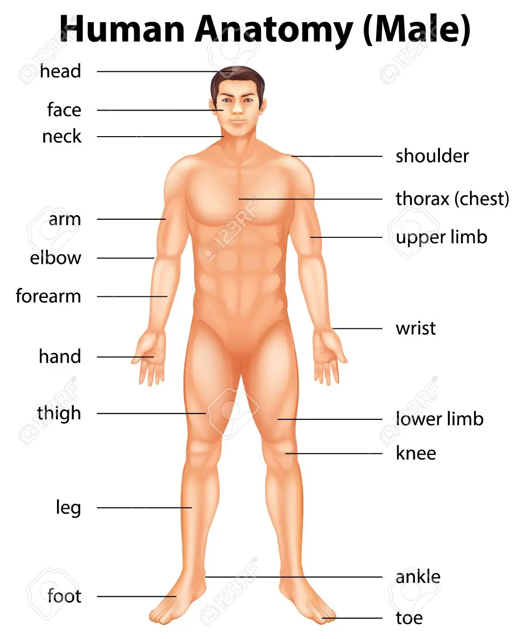
The human body is more like an entire factory, which produce from the rawmaterialsin the environment, what we need to keep our strength and good health, grow and develop.To do this, the body has got different parts which are specialised in different and specific activities in the body system. All body parts, be itexternalor internal, cooperates with other parts to fulfil their duties of making the body function. Some parts, like thebrainandheart, function without you thinking about it.

Parts of Human Body
Parts of Human Body

Other parts, like your arms, hands, legs, and feet have to be told what to do. Knowledge of our body parts and their location is very important for if you know a little about these human body parts, you are most likely to understand your health and theailmentsthat you suffer from even better.

This will help when seeking help while prompted by the body. It will also allow the person to keep their body healthy. Furthermore, having this knowledge is what separates humans from any other thing on the planet. We have the know-how on when and how to seek treatment if we would just listen to our bodies.